| McSwiggen & Associates |
 |
 |
 |
 |
 |
 |
 |
 |
| /Tech Notes/Map Analysis |

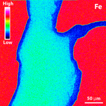
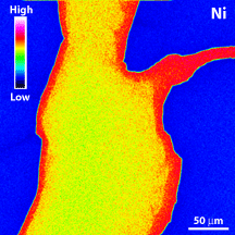
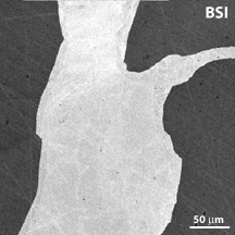
Map analysis allows the user to record the distribution of elements within an area of the sample. The mapped area can be tens of microns across or many centimeters across. There are two basic mapping modes that can be used. Either the beam can be scanned across the sample or the sample can be moved back and forth under a fixed beam. The former is used when mapping a small area, and the latter is used for large areas. The user determines the area to be mapped, the number of pixels in the map, and the distance between each measurement location on the sample. Each spectrometer is set to the position of a single X-ray line, and the X-ray counts are then measured at each spot as the beam is being rastered, or the sample is being moved under the beam. The number of counts at each location is displayed as variations in intensity of the corresponding pixel on the map. The maps can be displayed either as grayscale (as is the backscattered electron image, BSI, to the right), or in color (as are the Ni and Fe maps, below). The color maps are typically displayed with the areas of low counts in shades of blue, and those with high counts in oranges and reds. The amount of times it takes to collect a map will depend on the parameters chosen. A 250 x 250 pixel map, using a 20 millisecond dwell time at each point, will take about twenty minutes to collect. However, a 500 x 500 pixel map, using a twenty millisecond dwell time will take about one hour and twenty minutes. Typically element maps are qualitative in nature because they are displaying the X-ray intensity at each pixel. The backgrounds are usually not subtracted, nor are the data corrected for matrix effects. They show the relative abundance of the elements mapped. The examples below are Fe and Ni maps of an exsolution lamellae from the Estherville meteorite. |
Figure 1. Backscattered electron image of a exsolution lamellae in the Estherville meteorite. |
|
area shown in Figure 1. |
area shown in Figure 1. |