| |
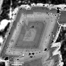
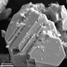
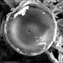
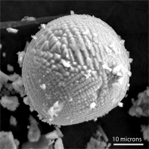
Electron microprobes have the same electron imaging capabilities as comparable scanning electron microscopes. Both backscattered electron and secondary electron images can be collected. Figures 1-4 are examples of both types of images. Backscattered electron images show compositional variations within the sample, whereas secondary electron images show variations in the surface morphology. For this reason, backscattered electron imaging is the most commonly used form of imaging on electron microprobes. Typically the sample going into an electron microprobe will be flat and polished, since this is required for good analyses. Because of the need for a smooth sample surface, secondary electron imaging will not provide much useful information. However, a backscattered electron image will allow the user to quickly distinguish grains of one phase from another, or to see compositional variations within a single grain. The backscattered electron image in Figure 1 shows an ankerite crystal that is zoned with varying amounts of iron and magnesium. The light areas are the areas with the higher backscattered electron signal and therefore must have a higher average atomic number. These are the regions higher in iron. |

Figure 1. Backscattered electron image of a zoned ankerite crystal.
|


